Segmenting X-Ray images with deep learning
STFC/Durham University CDT in Data Intensive Science.
Carolina Cuesta-Lázaro
Arnau Quera-Bofarull
Joseph Bullock
Who are we?


2 months PhD placement at IBEX innovations, as part of the CDT program
Carolina / Arnau
Cosmology
Joe
Particle Physics
Detect bone and soft-tissue on X-Ray images
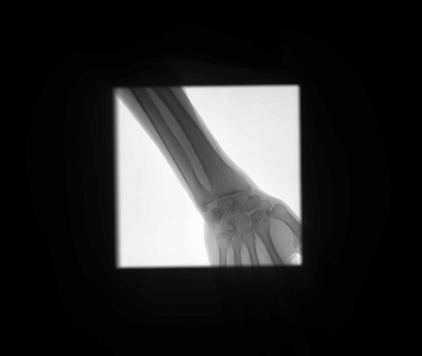
Detect
collimator
Segment
Open beam
Bone
Soft-tissue
SegNet
- The network has more than 15 Million free parameters.
- To find the values of the parameters that produce the correct segmentation, it has been trained on 1.3 Million images.

XNet
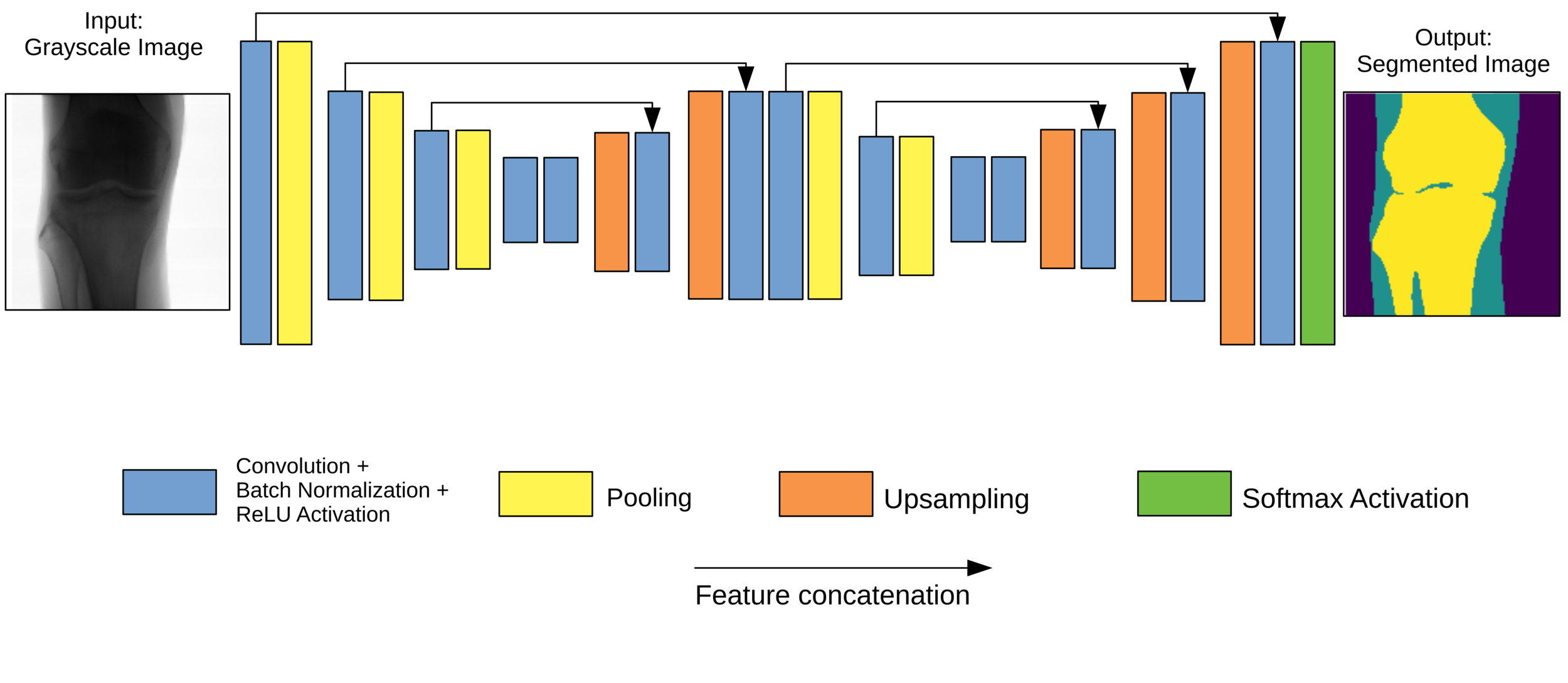
-
Original Architecture based on SegNet but fewer parameters.
- Trained on 150 images, artificially augmented to more than 7000.
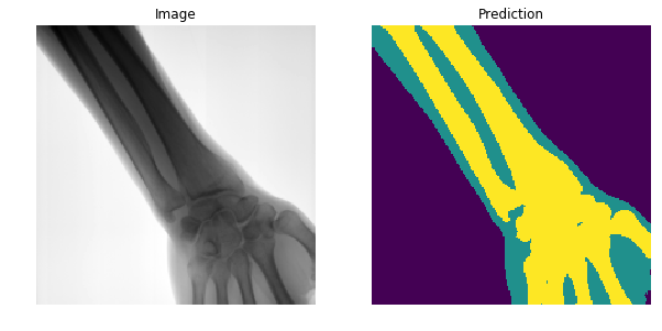
Results
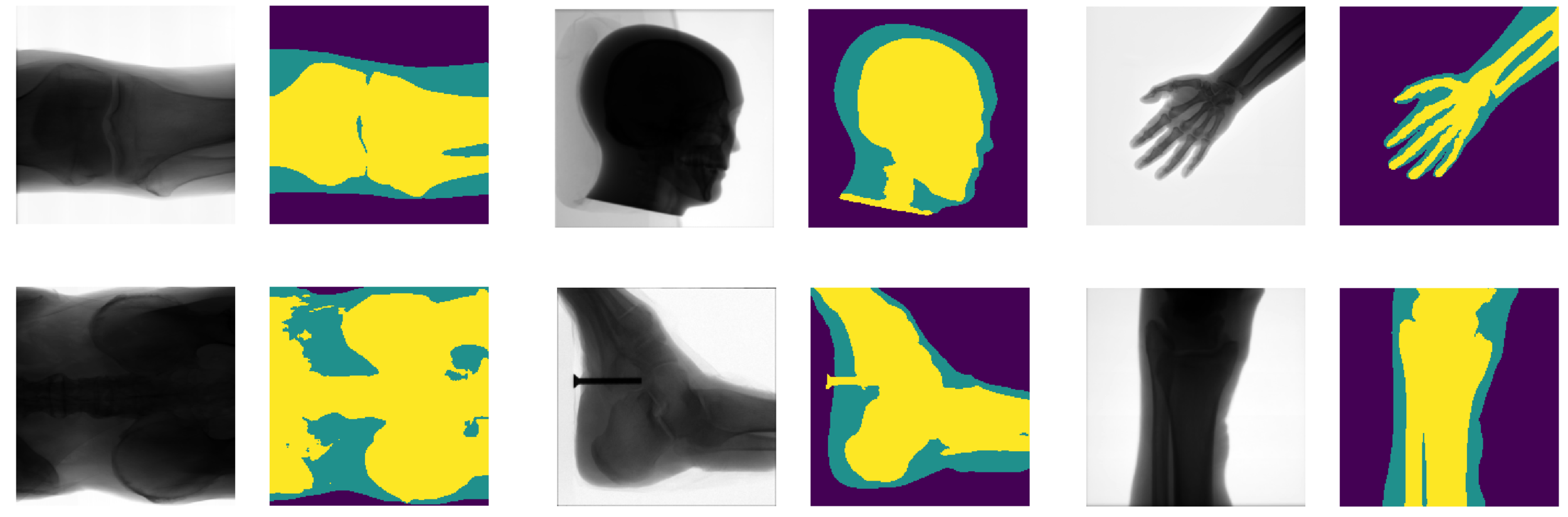
- Generalises well even for unseen categories!
- Overall accuracy on test set: 92%
- Soft tissue TP/FP rate: 82% / 4%
- Promising ML applications to medical imaging.
- Possible to train ML models with limited hardware and resources.
- Knowledge of building and deploying a machine learning product in an industrial setting.
- XNet Paper is on the arXiv:1812.00548v1, and will be presented in the upcoming SPIE Medical Imaging conference in San Diego.
Conclusions
Learning outcomes
NEPIC
By carol cuesta


